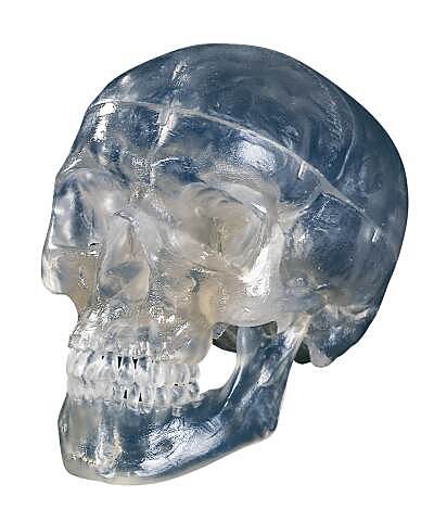
What if we were transparent?

We all know about sending sound waves through body tissues and analyzing the reflections with an ultrasound machine. The computer software and processor of the ultrasound machine then recreates a “picture” of what is inside. Ever wonder why can’t we just send light waves into a person and “see” what is inside in the same way?
“The reason a person is not transparent is that their tissues are highly scattering,” sending light waves careening through the tissue instead of straight through, as they would through the tissue of a jellyfish, explains Changhuei Yang of the California Institute of Technology. This light scattering, in addition to making us appear “solid”, makes the detection of our internal medical conditions considerably harder. We currently use a wide range of diagnostic tests and procedures to detect internal abnormalities. But Dr. Yang, an assistant professor of electrical engineering and bioengineering along with his colleagues at Caltech, MIT and in Switzerland have developed a method to counteract that scattering of light and remove the distortion it creates to potentially image our “insides”. If the technique can be developed in a practical manner, it may open up a whole new field of medical imaging and treatment.
Light scattering by a material is not exactly the random and unpredictable process one might imagine. In fact, scattering is deterministic, which means that the path that a beam of light takes as it traverses a particular slice of tissue and bounces and rebounds off of individual cells, is entirely predictable; if you again bounce a given source light through that same swath of cells, it will scatter in exactly the same way. If you can gather up the individual scattered photons of light and send them back through the tissue, they would bounce back through the specimen and converge at the exact spot from where they were sent.
“The process is similar to the scattering of billiard balls on a pool table. If you can precisely reverse the paths and velocities of the billiard balls, you can cause the billiard balls to reassemble themselves into a rack,” Yang explained. Dr. Yang, along with his colleagues at Caltech, École Polytechnique Fédérale de Lausanne in Switzerland, and MIT, exploited this phenomenon to offset the opaque nature of our tissues.
The technique, called turbidity suppression by optical phase conjugation (TSOPC), is done by using a holographic crystal to record the scattered light pattern emerging from a 0.46-mm-thick piece of chicken breast. The researchers then send the holographic pattern collected back through the tissue section to recover the original light beam.
“This is similar to grabbing hold of the direction of time flow and turning it around; the time-reversed photons must retrace their trajectories through the tissue,” explained Dr. Yang. “The task is formidable though, as this is comparable to starting with a rack of 10 to the 18th power billiard balls (or photons), scattering them around the table, and attempting to reassemble them into a rack.”
I am reminded of a discussion given by that amazing scientist and thinker, Dr. Richard Feynman in his 1983 video where he points out that you could determine who and when a person dived into a swimming pool by analyzing the waves in the pool generated by that person. All of imaging in medicine really boils down to being able to sort out the patterns of the waveforms. The following video is 5 or 6 minutes long, but is well worth watching to begin to understand the concepts that Dr. Yang and his colleagues are using. (Click here or on picture to view the video)
There are numerous potential applications for this technique, not just for imaging, but for treatment. Imagine being able to collect the light scatter data from a tissue which shows the outline of a tumor. The hologram image you collect from sending a light into the tissue contains information about the path that the scattered light traveled when it was sent through the tissue. That hologram would also describe the optimal path back to the tumor tissue.
Currently a whole range of photosensitizer drugs is available for photodynamic therapy. A number of these compounds can attach to cancer cells. If a photosensitizing agent is injected into the bloodstream the agent is absorbed by cells all over the body, but stays in cancer cells longer than it does in normal cells. Approximately 24 to 72 hours after injection, most of the agent has left normal cells but remains in cancer cells. If the tumor is then exposed to light, the photosensitizer in the tumor absorbs the light energy and produces an active form of oxygen that destroys nearby cancer cells. Using the holographic data to send light in reverse fashion, may be able to very precisely destroy malignant cells and leave normal body cells unharmed.
Another potential application of this new technique could offer a way to power miniature implants buried deep within tissues. Photovoltaic batteries power spacecraft on Mars. What if you could use the technique to power a cardiac pacemaker by shining a special holographic light power source through the tissues to a photovoltaic battery to charge up and power the microprocessor? Right now, cardiac pacemakers need to be the size they are largely because the implantable batteries must last several years (and they always need to be replaced sooner or later). A much smaller battery (and pacemaker) could be engineered if it were possible to charge up an implanted photovoltaic battery on a regular basis with a holographic light beam! Think of pacemakers the size of a dime, or a pencil eraser..
